Visualizing fungal infections deep in living host tissue reveals proline metabolism facilitates virulence

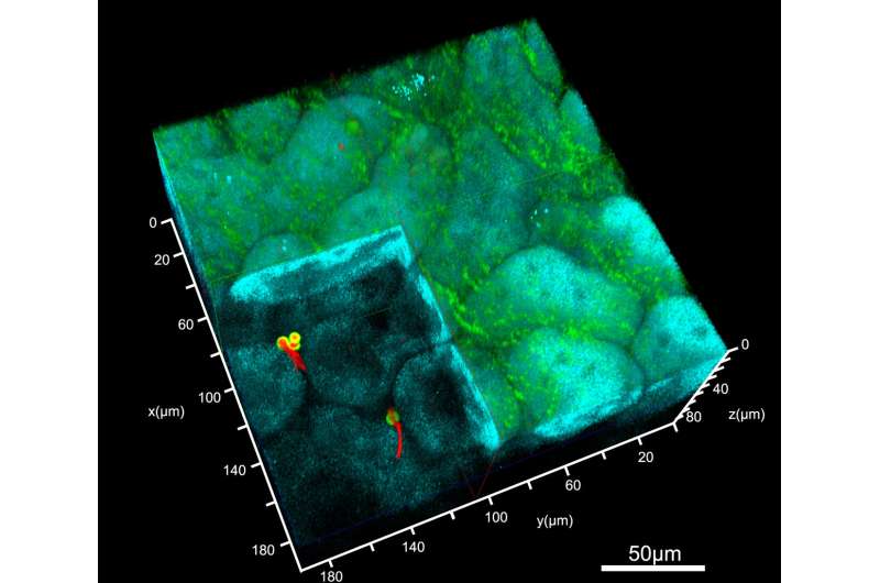
An international team of scientists, led by researchers from the Department of Molecular Biosciences, The Wenner-Gren Institute, SciLifeLab, Stockholm University has published in PLoS Pathogens the first successful application of two-photon intravital microscopy (IVM) to image the dynamics of fungal infections in the kidney of a living host. The study reveals that the opportunistic human fungal pathogen Candida albicans requires the ability to metabolize proline, an amino acid obtained from the host, to mount virulent infections.
Candida albicans has recently been listed as one of four “critical priority” fungal pathogens by the World Health Organization (WHO). A defining feature of C. albicans is that it is a commensal organism that thrives in symbiosis along with other components of the human microflora, and is normally well-tolerated. However, when humans experience health challenges that negatively affect the immune system, C. albicans can cause bloodstream infections that are lethal unless aggressively treated.
During infection, C. albicans cells are known to switch morphologies from ovoid yeast-like to elongated filamentous hyphal cells, a process linked to the capacity of this fungi to grow as a pathogen. Although proline has long been known to trigger hyphal growth, it has been recently discovered that C. albicans can use proline as a main source of energy. Proline breakdown takes place in the mitochondria, the powerhouse of the cell.
The investigating team now reports that proline is an important source of energy in other pathogenic Candida species, including the multidrug resistant C. auris, an emerging health threat and also a WHO critical priority fungal pathogen. The lead author of this study, Dr. Fitz Gerald S. Silao, explains, “In fungal cells possessing mitochondria equipped with a full complement of energy conserving respiratory complexes, the catabolism of proline generates almost as much chemical energy (ATP) as does the catabolism of the energy-rich sugar glucose.”
The collaborating groups provided access to a multitude of infection models, including artificial skin, co-culture with immune cells, survival in whole human blood and two model host systems. The results consistently showed that strains unable to metabolize proline exhibit significantly reduced virulence properties and a clearly diminished capacity to undergo morphological transitions. These observations provide novel insights implicating proline metabolism as a key determinant of pathogenic fungal growth.
Proline, one of the 20 naturally occurring amino acids in the body, is enriched in extracellular matrix proteins such as collagen, and as such is readily available when connective tissue is broken down at sites of infection, or when a host becomes vulnerable as a result of cancerous growth or upon the onset of sarcopenia. Strikingly, genetic dissection of the Proline UTilization (PUT) pathway and of the control mechanisms governing proline use led to the discovery that proline is toxic to cells unable to catabolize it. This latter finding was unexpected, and further work is needed to solve the mystery and underlying mechanism of its toxicity.
The kidney is the primary organ affected during bloodstream infections by C. albicans, and it is imperative to understand why. To obtain answers, the team initially applied a mouse model of infection, which remains invaluable for this kind of work, and found that C. albicans cells lacking the capacity to utilize proline were less invasive and virulent. Notably, mice infected with fungal cells unable to catabolize proline, e.g., cells lacking the Put2 enzyme (1-pyrroline-5-carboxylate (P5C) dehydrogenase), showed milder illness or no symptoms at all.
Next, state-of-the-art 2-photon intravital microscopy (IVM) was used to visualize, in real-time, the invasion of C. albicans cells deep in the cortex of the kidneys inside a living host. In contrast to native wild-type cells, the Put2-deficient cells (put2-/-) failed to form hyphae in kidneys. It should be recognized that imaging the initial stages of an ongoing infection is very challenging due to many factors, such as the small size and scarcity of the fungal cells and the continuous motions associated with vital and life-sustaining processes in living hosts.
“The utilization of engineered reporter strains and differential staining techniques enabled us to swiftly spot fungal outgrowth deep in the tissue. IVM is really a game-changer as it allows the imaging of a dynamic process in organs in their intact physiological context and at depths unachievable with conventional fluorescence or confocal microscopy,” says Dr. Christiane Peuckert, Head of the IntraVital Microscopy Facility-Stockholm University (IVMSU) and co-corresponding author of the paper.
As the senior author of this study Professor Per O. Ljungdahl explains, “The kidney is a major hub for proline metabolism, which makes our data congruent to known processes linked to kidney function and make this work even more interesting. Our future research will focus on creating sets of reporter strains to directly assess and visualize proline utilization in the kidney. We will also extend the application of IVM to other Candida species to determine whether a tailored proline metabolic network tuned to the mammalian host environment is a common and key feature of important opportunistic human fungal pathogens that are of growing concern to human health.”
More information:
Fitz Gerald S. Silao et al, Proline catabolism is a key factor facilitating Candida albicans pathogenicity, PLoS Pathogens (2023). DOI: 10.1371/journal.ppat.1011677
Citation:
Visualizing fungal infections deep in living host tissue reveals proline metabolism facilitates virulence (2023, November 2)
retrieved 2 November 2023
from https://phys.org/news/2023-11-visualizing-fungal-infections-deep-host.html
This document is subject to copyright. Apart from any fair dealing for the purpose of private study or research, no
part may be reproduced without the written permission. The content is provided for information purposes only.
