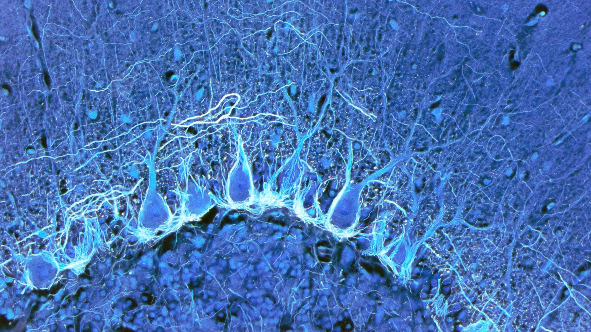
Scientists just unveiled the largest and most detailed “atlas” of the human brain ever created.
It details the arrangement and inner workings of 3,300 types of brain cells, only a fraction of which were previously known to science. The research was released Thursday (Oct. 12) in the form of 21 new papers published across three journals: Science, Science Advances and Science Translational Medicine.
“It’s not just an atlas,” Ed Lein, a neuroscientist at the Allen Institute for Brain Science and a lead author of five of the papers, told MIT Technology Review. “It’s really opening up a whole new field, where you can now look with extremely high cellular resolution in brains of species where this typically hasn’t been possible in the past.”
The research was conducted as part of a National Institutes of Health project known as the Brain Research through Advancing Innovative Neurotechnologies (BRAIN) Initiative Cell Census Network, or BICCN. Launched in 2017, the massive project aims to catalog the cells found in the brains of mice, humans and nonhuman primates such as monkeys.
Related: We finally know why the brain uses so much energy
These cells include neurons, the brain cells that communicate through chemical and electrical messages, and a roughly equal number of non-neuronal cells. These non-neuronal cells include glia, a class of brain cells that provide structural support, nutrients and insulation to neurons while also regulating how they send signals. The adult human brain contains an estimated 86 billion neurons, give or take about 8 billion, and another 84 billion or so of these non-neuronal cells.
The BICCN human brain atlas used cutting-edge techniques that had previously mostly been used in animals, Mattia Maroso, a senior editor for the journal Science, wrote in the special issue published Thursday.
Scientists used transcriptomics, which involves cataloging all the RNA in individual cells; RNA is a genetic molecule that contains instructions to build proteins and does other important jobs. They also used epigenomics, which involves examining chemical tags that sit on top of DNA and control how genes can be used. Single studies included in the BICCN included data from hundreds of thousands to millions of brain cells.
Combining these techniques, researchers created single-cell-scale maps of the developing and adult human brain, as well as the brains of primates called marmosets (Callithrix) and macaques (Macaca). Some studies also looked at the brains of chimpanzees (Pan troglodytes) and gorillas (Gorilla).
This allowed for a direct comparison between human and nonhuman primate brains, and revealed that the myriad cell types found in our brains are also found in chimps and gorillas, The New York Times reported. But although we share cell types, the gene activity of those cells appears to be markedly different in humans and apes, and this changes how those cells work together.
“It’s really the connections — how these cells are talking to each other — that makes us different from the chimpanzees,” Trygve Bakken, a neuroscientist at the Allen Institute who worked on the primate studies, told the Times.
Although unprecedented in its detail, the human brain atlas is a first draft. In the future, scientists want to decode the function of the newly discovered cells in the brain, many of which lie deep in the brain, in structures like the brain stem, Nature reported. They also want to understand how the gene activity of different cells contributes to the development of neurological diseases.
In a statement that ran alongside the 21 newly published papers, Science said, “If we want to understand what makes us human, and the mechanisms responsible for the development of neurological disorders, we first need to have a deep knowledge of the human brain at the cellular level, which is exactly what this collection of papers from the BICCN is about.”